Quick Summary
Click here for Price and Turnaround Time
Phenotype: Progressive retinal atrophy is characterized by bilateral degeneration of the retina resulting in progressive vision loss leading to total blindness. Clinical signs of PRA1 appear around 6 years of age. Clinical symptoms of PRA2 appear around 4 years of age.
Mode of Inheritance: Autosomal recessive
Alleles:
N = Normal
PRA1 = Golden Retriever progressive retinal atrophy (variant 1)
PRA2 = Golden Retriever progressive retinal atrophy (variant 2)
Breeds appropriate for testing: Golden Retriever, Golden Doodle, Labrador Retriever
Explanation of Results:
- Dogs with N/N genotype are not expected to develop either of these two forms of Golden Retriever progressive retinal atrophy and cannot transmit these progressive retinal atrophy variants to their offspring.
- Dogs with N/PRA1 or N/PRA2 genotype are not expected to develop either of these two forms of Golden Retriever progressive retinal atrophy, but are carriers. They may transmit a Golden Retriever progressive retinal atrophy variant to 50% of their offspring. Matings between two carriers of the same variant (e.g. N/GR_PRA1 x N/GR_PRA1) are predicted to produce 25% of puppies affected by that form of progressive retinal atrophy. Matings between two carriers of the different variants (N/GR_PRA1 x N/GR_PRA2) will not produce affected offspring.
- Dogs with PRA1/PRA1 or PRA2/PRA2 are expected to develop progressive retinal atrophy. They will transmit respective progressive retinal atrophy variants to all of their offspring.
Results of this test can be submitted to the OFA (Orthopedic Foundation for Animals)
Golden Retriever Health Panel
$135 per animal
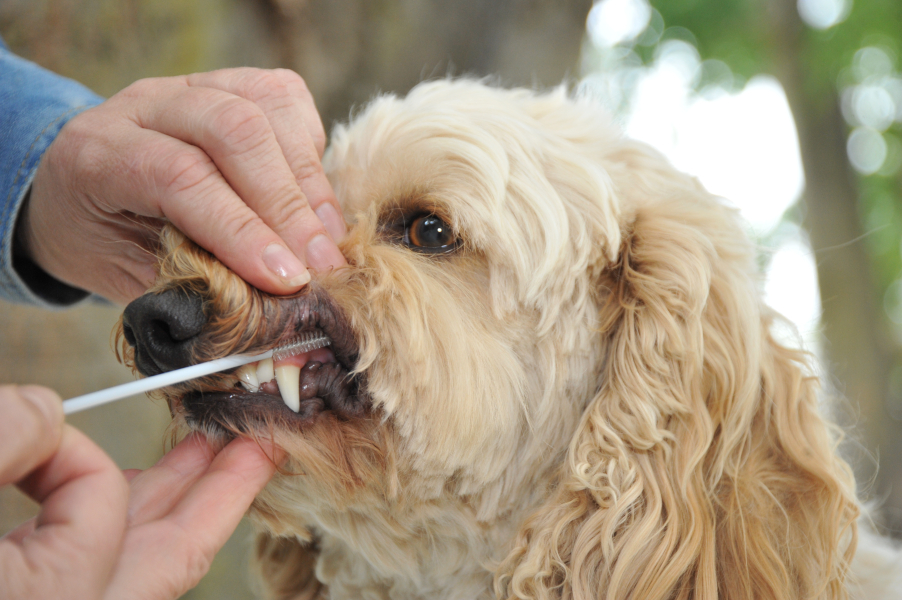
Sample Collection
Dog DNA tests are carried out using cells brushed from your dog's cheeks and gums. The preferred cytology brushes are sent to you by mail, or you may provide your own brushes. For accepted alternative brushes, click here
We recommend waiting until puppies are at least three weeks old before testing.
Step-By-Step:
- Make sure the dog has not had anything to eat or drink for at least 1 hour prior to collecting sample.
- When swabbing puppies, isolate each puppy from the mother, littermates and any shared toys for 1 hour prior to swabbing. Puppies should not have nursed or eaten for 1 hour prior to collecting sample.
- If collecting samples from more than one dog, make sure to sample one dog at a time and wash your hands before swabbing another dog.
- Label brush sleeve with name or ID of dog to be sampled.
- Open brush sleeve by arrow and remove one brush by its handle.
- Place bristle head between the dog’s gums and cheek and press lightly on the outside of the cheek while rubbing or rotating the brush back and forth for 15 seconds.
- Wave the brush in the air for 20 seconds to air dry.
- Insert brush back into sleeve.
- Repeat steps 5 - 8 for each unused brush in sleeve on a fresh area of cheek and gums. Make sure to use and return all brushes sent by the VGL. In most cases, it will be 3 brushes per dog. If using interdental gum brushes, please note that the VGL requires 4 brushes per dog and only moderate or wide interdental gum brushes are accepted.
- Do not seal brushes in sleeve.
- Place all samples in an envelope and return to the address provided.
ATTENTION:
- Do not collect saliva/drool – the key to obtaining a good sample is getting cheek cells on the swab
- Do not rub swab on the dog’s tongue or teeth – this will result in poor quality sample
- Do not collect a sample from a puppy that has recently nursed – the mother’s genetic material can rub off on the puppy’s mouth and contaminate the sample
More than 20 mutations have been identified that result in canine progressive retinal atrophy (PRA). The condition is characterized by bilateral degeneration of the retina resulting in progressive vision loss leading to total blindness. Golden Retrievers are affected by more than one form of PRA with mutations in three distinct genes having been identified. Two of such mutations are known as PRA1 and PRA2. PRA1 results from a mutation in the SLC4A3 gene and accounts for over 60% of diagnosed Golden Retrievers. PRA2 results from a mutation in the TTC8 gene and accounts for 30% of Golden Retrievers diagnosed with PRA. Both mutations are autosomal recessive thus two copies of the same affected gene must be present for the disease to be observed and both males and females are equally affected. Presence of one copy of each affected genes in the same dog will not cause blindness. Clinical signs of PRA1 appear around 6 years of age. Clinical symptoms of PRA2 appear around 4 years of age. Breeding two carriers with the same mutation is predicted to produce 25% affected puppies and 50% carriers. Mating a carrier of PRA1 (that is N/N for PRA2) with a carrier of PRA2 (that is N/N for PRA1) will not produce affected animals.
Testing for PRA1 and 2 assists owners and breeders in identifying affected and carrier dogs. Breeders can use results from the tests as a tool for selection of mating pairs to avoid producing affected dogs. Genetic testing for PRA1 and 2 is recommended for Golden Retrievers and Golden Doodles. PRA2 has been reported at low frequency in Labrador Retrievers, but an extensive survey of the US population has yet to be investigated.