Quick Summary
Click here for Price and Turnaround Time
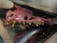
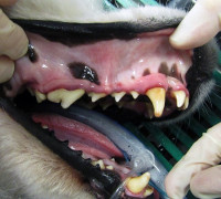
Phenotype: Familial enamel hypoplasia (FEH) is characterized by enamel pitting and tooth discoloration. FEH in Samoyeds is usually noticed when the permanent teeth erupt, around 5-6 months of age. The deciduous teeth appear normal. The disorder is characterized by marked thinning or absence of the outer enamel layer with irregular pitting, which predisposes the dog to patchy tooth discoloration, dentin sensitivity, premature tooth wear, dental caries, pulp exposure, tartar accumulation, gingivitis, and periodontitis. Periodontal disease can lead to bone loss, gum recession, and tooth loss. Halitosis (foul-smelling breath) is a common effect of the dental disease.
Mode of Inheritance: Autosomal recessive
Alleles: N = Normal, FEH = Familial enamel hypoplasia
Breeds appropriate for testing: Samoyed
Explanation of Results:
- Dogs with N/N genotype will not have familial enamel hypoplasia and cannot transmit this variant to their offspring.
- Dogs with N/FEH genotype will not be affected by familial enamel hypoplasia, but are carriers. They will transmit this variant to 50% of their offspring. Matings between two carriers are predicted to produce 25% familial enamel hypoplasia-affected puppies.
- Dogs with FEH/FEH genotype will have familial enamel hypoplasia and will transmit this variant to all of their offspring.
Results of this test can be submitted to the OFA (Orthopedic Foundation for Animals)
Sample Collection
Dog DNA tests are carried out using cells brushed from your dog's cheeks and gums. The preferred cytology brushes are sent to you by mail, or you may provide your own brushes. For accepted alternative brushes, click here
We recommend waiting until puppies are at least three weeks old before testing.
Step-By-Step:
- Make sure the dog has not had anything to eat or drink for at least 1 hour prior to collecting sample.
- When swabbing puppies, isolate each puppy from the mother, littermates and any shared toys for 1 hour prior to swabbing. Puppies should not have nursed or eaten for 1 hour prior to collecting sample.
- If collecting samples from more than one dog, make sure to sample one dog at a time and wash your hands before swabbing another dog.
- Label brush sleeve with name or ID of dog to be sampled.
- Open brush sleeve by arrow and remove one brush by its handle.
- Place bristle head between the dog’s gums and cheek and press lightly on the outside of the cheek while rubbing or rotating the brush back and forth for 15 seconds.
- Wave the brush in the air for 20 seconds to air dry.
- Insert brush back into sleeve.
- Repeat steps 5 - 8 for each unused brush in sleeve on a fresh area of cheek and gums. Make sure to use and return all brushes sent by the VGL. In most cases, it will be 3 brushes per dog. If using interdental gum brushes, please note that the VGL requires 4 brushes per dog and only moderate or wide interdental gum brushes are accepted.
- Do not seal brushes in sleeve.
- Place all samples in an envelope and return to the address provided.
ATTENTION:
- Do not collect saliva/drool – the key to obtaining a good sample is getting cheek cells on the swab
- Do not rub swab on the dog’s tongue or teeth – this will result in poor quality sample
- Do not collect a sample from a puppy that has recently nursed – the mother’s genetic material can rub off on the puppy’s mouth and contaminate the sample
Autosomal recessive amelogenesis imperfecta (ARAI) is an inherited genetic disorder of tooth enamel that occurs in humans. It is commonly known as familial enamel hypoplasia (FEH) in dogs. Dr. Niels Pedersen and his research group at the School of Veterinary Medicine, University of California, Davis have researched FEH in several breeds and have identified breed-specific mutations in two breeds, the Italian Greyhound and the Samoyed. Mutations in other breeds have yet to be identified.
The common occurrence of FEH in pure breeds of dogs and ARAI in humans is related to the complexity of tooth formation. The formation of enamel is the last step in tooth development and involves the activity of many genes and their proteins acting at the same or different times in pre- and post-natal life. The involvement of many genes increases the likelihood of a deleterious mutation occurring. These mutations are inadvertently subjected to positive selection in association (linkage) with traits that are deemed desirable, frequently from a popular sire.
Amelogenesis imperfecta in humans can be autosomal dominant or recessive, or sex-linked. The mutation can be associated only with the teeth (non-syndromic) or can involve several structures in the body (syndromic). FEH in dogs appears to be predominantly non-syndromic, autosomal recessive in inheritance, and the causative mutation unique to each breed. The autosomal recessive mutation in Samoyed responsible for FEH has been confirmed. An analogous mutation has been associated with a specific form of ARAI in humans. Based on preliminary testing, it is estimated that 1-2% of Samoyed are affected with FEH and over 10% of dogs with normal-appearing teeth are carriers.
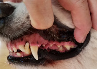
FEH in Samoyed is usually noticed when the permanent teeth erupt, around 5-6 months of age. The deciduous teeth appear normal. The disorder is characterized by marked thinning or absence of the outer enamel layer with irregular pitting, which predisposes to patchy tooth discoloration, dentin sensitivity, premature tooth wear, dental caries, pulp exposure, tartar accumulation, gingivitis, and periodontitis. Periodontal disease can lead to bone loss, gum recession, and tooth loss. Halitosis is a common sequela of the dental disease.
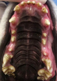
Figure 1 shows a Samoyed with normal teeth. Figures 2 and 3 show typical Samoyed with FEH, and figure 4 shows the results of restorative and cosmetic dentistry.
Testing for FEH assists owners and breeders in identifying affected and carrier dogs. Breeders can use results from the test as a tool for selection of mating pairs to avoid producing affected dogs.